Nanillænus americanus (BILLINGS 1859)
A not so common, but no so rare, trilobite from the USA. It is rarely found complete, generally found in the form of cephala or pygidia. A classic location for this trilobite is the Trenton Group, at West Canada Creek, New York.
| 1859 |
Illænus americanus |
nov. sp. BILLINGS 1859, p. 371. |
| 1865 |
Illænus americanus |
BILLINGS 1859 p. 329, figs. 316ad, 318. |
| 1908 |
Illænus americanus |
RAYMOND & NARRAWAY, pl. 60, figs. 1-3. |
| 1947 |
Illænus americanus |
WILSON p. 31, pl. 7, figs. 3, 4. |
| 1979 |
Nanillænus americanus |
LUDVIGSEN, 1979a, p. 37 |
| 1999 |
Nanillænus americanus |
BRETT et al., p. 299, figs. 7.6, 7. |
| 2002 |
Nanillænus americanus |
WHITELEY et al., p. 122, pl. 8. |
NB : The type for this species is lost (Wilson, 1947, p. 31).
|
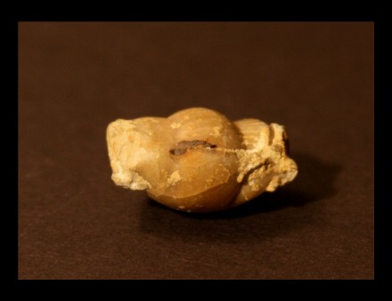
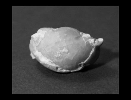
Specimen description
- Enrolled trilobite..
- Free of matrix.
- Distinct left eye. Right eye partially prepared
- Thoracic rings perfectly preserved.
- Total size : 31 mm (width : 16 mm)
- Upper Ordovician.
- Minnesota.
- USA.
|
|
Dorsal view of the cephalon |
Dorsal view of the pygidium |
DIAGNOSIS
|
Order
|
Corynexochida
|
KOBAYASHI 1935 |
Occurrence: Lower Cambrian Middle Devonian
- Cephalon: Opisthoparian sutures.
- Glabella elongate, sides often concave (pestle-shaped).
- Furrows (when not effaced) typically with splayed arrangement, the
hind pair pointing sharply backwards. Anterior pairs tending more and
more forward directed. Sometimes furrows pit-like.
- Cranidial borders often ledgelike.
- Hypostome conterminant or (in derived forms) impendent.
- Eyes typically large, in some gently arcuate
- Thorax: typically with 7-8 segments (but range for order is
2-12, rarely more).
- Pleural tips often spinose.
- Pygidium : typically large (isopygous or subisopygous).
- Variable form.
- Some spinose.
|
|
Suborder
|
Illænina
|
JAANUSSON 1959 |
Occurence : Upper Cambrian Devonian.
- Cephalon : Typically effaced.
- Doublure broad.
- Opisthoparian facial sutures.
- Sutures distinctly diverging anteriorly.
- Glabella expanding forwards.
- Lateral furrows often faint or absent.
- 4 pairs of glabellar furrows, commonly faint or absent.
- Extra-axial cephalic muscle impressions (lunette) present.
- Eyes frequently on the posterior part of the genæ, close to
the axial furrows.
- Terrace lines frequent, particularly at the distal ends of the thoracic
exoskeleton and the ventral doublure.
- Hypostome impendent (conterminant, but hypostome no longer matching
anterior glabellar border)
- Rostral plate limited by sutures, fused in the late species of Panderia.
- Trilobitation frequently fainting .
- Thorax : 8-10 segments.
- Pygidium : Isopygous or subisopygous.
- Rounded posteriorly.
- Usually with short axis.
|
|
Superfamily
|
Illænoidea |
HAWLE & CORDA
1847 |
|
| Family |
Illænidæ |
HAWLE &
CORDA 1847 |
- Cephalon : Axial region of cephalon smooth, merging forward
into frontal area without boundary.
- 4 pairs of muscle scars can be observed instead of glabellar and occipital
furrows.
- Glabella strongly convex.
- Opisthoparian facial sutures.
- Rostral shield broad (tr.).
- Hypostoma oval, with elongated body (sag.)
- Anterior wings of hypostoma broad, quadrangular.
- Pygidium : Axis and pleural field smooth or with very faint
traces of unfurrowed ribs.
- Surface ornemented with terrace lines or small pits, or both.
- No tuberculate or granulose ornementation.
|
| Genus |
Nanillænus |
JAANUSSEN 1954 |
|
| Species |
americanus |
(BILLINGS 1859) |
Nanillaenus species oblong and distinctly trilobed.
- Cephalon : Significantly larger than the pygidium and has a length two thirds the width.
- Most prominently convex at the center.
- Eyes set well back and far apart.
- Free cheeks with a prominent blunt angle.
- Thorax :8 thoracic segments
- Aaxial lobe about one third the width of the whole trilobite.
- Pygidium : Relatively small, having a length two thirds that of the cephalon and a width twice that of the length.
- Strong axial furrows that are well defined over half the length of the pygidium which continue through the thorax and end on the cephalon as sigmoid curves (curving in then out).
- Surface is covered with short squamose (“resembling a scale”) wrinkles.
|
Complementary description :
- Cephalon : well-developed eyes.
- Lunettes at the anterior end of the cephalic axial furrows.
- Significantly deep axial furrows.
- Long, hour-glass shaped, rostral flange.
- Pygidium : significantly deep axial furrows.
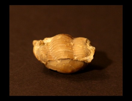
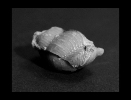
Complementary pictures :
 |
 |
 |
Oblique view of the thorax and the left eye. |
Oblique view of the thorax and the right eye. |
Dorsal view of the cephalon. |
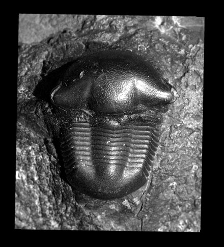
Other specimen found on internet :
Back to
Home Page