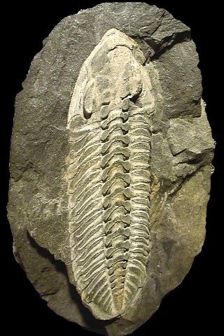
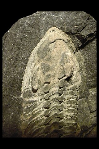
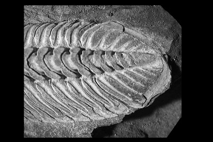
Here a second specimen of this European zonal trilobite, definitely better in quality than the Ph. micheli micheli from Brittany presented, very compressed laterally (traditional mode of preservation on the site of Santa Justa) but which nevertheless preserved a number of details of which in particular the occular facets, remarkably visible at the right eye.
The trilobites of Valongo (Portugal) often are very coloured and this trilobite does not escape the rule: silver plated white contrasting with a gray blue matrix.
| Order
|
Phacopida
|
SALTER 1864
|
Occurrence: Lower Ordovician
(Tremadoc) to Upper Devonian (Famennian).
- Cephalon: Proparian (Phacopina and Cheirurina), gonatoparian
(Calymenina) or opisthoparian (Calymenina),
- Preglabellar field often very short or absent.
- 4 or fewer pairs of glabellar furrows.
- Eyes : when present, schizochroal (Phacopina) or holochroal (Cheirurina
and Calymenina).
- With rostral plates (Calymenina and Cheirurina) or without (some
Phacopina).
- Hypostome conterminant (all suborders) to impendent (some devonian
Phacopina).
- Exosqueletton generally granulous.
- Thorax: 8 19 segments, sometimes distinctly furrowed.
- Axis sometimes broad (e.g., Homalonotidae).
- Pygidium : Typically micropygous (most Calymenina and Phacopina),
but variable (e.g., subisopygous in Dalmanitoidea and Acastoidea).
- May be lobed or spiny (e.g., Cheirurina, some Dalmanitoidea, Acastoidea),
or smooth-margined, with round or subtriangular outline (e.g., Calymenina,
Phacopoidea).
|
| Sub-order
|
Phacopina
|
STRUVE
1959
|
- Cephalon : Proparian sutures (sometimes fused).
- Schizochroal eyes.
- Glabella expands forwards.
- Librigena typically yoked as single piece.
- Hypostome conterminant to impendent, some with no rostral plate.
- Some with genal spines.
- Thorax: 10 to (typically) 11 segments.
- Pleurae furrowed, articulating facets distinct.
- Rounded, angular, or spinose tips.
- Pygidium : Typically smaller than cephalon (but subisopygous
in Dalmanitoidea and Acastoidea).
- Smooth or spinose.
|
| Superfamily
|
Phacopoidea
|
HAWLE
& CORDA 1847
|
- Cephalon : Auxiliary impressions short or lacking.
- Dorsal furrows strongly diverging.
- "anterior" lobe composite.
- No genal spines.
- Vincular furrow generally present.
|
| Family
|
Phacopidæ
|
HAWLE & CORDA
1847
|
- Cephalon : Glabella broadening markedly forward.
- S2 and S3 lateral glabellar furrows obsolescent.
- S1 generally transglabellar, thus forming a more or less distinct
"intercalating ring".
- Auxiliary impressions field ovale to circular.
- Genal angles rounded, lobiform or angular.
- No genal spines.
- Hypostoma triangular to trapezoidal.
- Thorax : Thoracic pleurae with rounded ends.
- Pygidium : Well-rounded, semicircular or shorter.
- Margin complete.
- No spine.
|
Genus |
Phacopidina |
BANCROFT 1949 |
- Cephalon : Ogival.
- Anterior cephalic border distinct, more or less flatenned.
-
Suture slightly isolated of the furrow préglabellaire.
-
Frontal lobe losangic.
-
Glabellar furrows S2 and S3 less marked than the deep and forked furrow S1.
-
Very short genal points.
-
Vincular furrows on each lateral side of the doublure.
-
Pygidium: Small, triangular.
-
Rachis with 5 to 8 rings.
-
Pleural lobes prolonged by a more or less developed terminal spine, incised by 4 to 5 pairs of pleural ribs.
|
Species |
micheli |
TROMELIN 1877
|
- Cephalon: Glabella non claviform, limited by narrow dorsal furrows.
- Occipital furrow slightly marked in its median part.
- Tiny genal points.
- Lateral border of the genae punts with more or less sharp edges.
- Lateral cephalic furrows marked well at the level of the eyes.
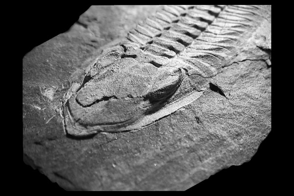
- Very large kidney-like eyes comprising 28 or 29 dorso-ventral files with nine lenses to the maximum.
- Narrow cephalic doublure.
- Very lengthened hypostom, with posterior border strip-like equipped with 2 small round outgrowths.
- Pygidium: 7 or 8 axial rings.
- 4 pairs of pleural ribs with obsolete interpleural furrows.
- Tiny hardly visible terminal spine.
|