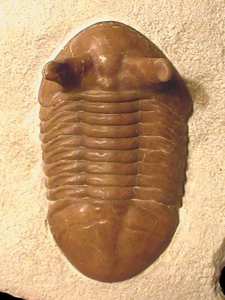
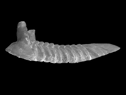
This is a trilobite occuring in the Lower Ordovician, Llanvirn, of the North-West
of Russia. This is one of the 35+ species of the Asaphus genus which
nicest specimens are collected and prepared by and at the Paleolab
of St Petersburg. Historically, most of the Asaphids have been found and described
in Northern Europe, but the exceptional preservation of the russian specimens
lead many collectors to renew their former specimens.
|
Order
|
Asaphida
|
SALTER 1864
|
Order including 1/5 of all the species of Trilobites,
it mostly regroup librostoms of various morphology, in which the most advanced
families do present a ventral median suture early visible in the ontogeny
( "asaphoïd" protaspid larvæ).
Occurrence : Middle-Upper Cambrian boundary to upper Ordovician-lower
Silurian.
- Cephalon : Opisthoparian.
- Often equal / subequal to pygidium (e.g., Asaphoidea), but some not
so (e.g., Trinucleioidea).
- Usually with a high degree of cephalic effacement so glabellar furrows
are faint or not visible.
- Eyes usually large (some forms secondarily blind).
- Preoccipital glabellar tubercle in late forms.
- Cephalic doublure often wide, with terrace ridges.
- Librigenæ are typically separated by a median ventral suture.
- Dorsal anterior facial sutures often curve adaxially to meet in front
of the glabella.
- Hypostome conterminent or impendent, with only primitive forms (e.g.,
the Anomocaroidea) natant.
- Thorax : Typically 5 12 segments, but 2 - 3 in a few Trinucleioidea,
13+ in some Anomocaroidea, up to 30 in an Alsataspidid (Trinucleioidea).
- Pygidium: Typically large (subisopygous to macropygous).
- Wide doublure.
|
|
Superfamily
|
Asaphoidea
|
BURMEISTER 1843
|
Occurrence: Middle Cambrian to Ordovician.
- Cephalon : Preoccipital glabellar tubercle.
- Glabella elongate, subparallel to tapering forward.
- Defined occipital ring.
- Curved, apostrophe-like pair of basal glabellar furrows isolated within
glabella.
- Hypostome conterminant, fixed to the doublure (rarely impendent)
- Thorax : 6 - 9 segments, typically 8.
- Pygidium : Typically rounded.
- Typically without spines, sometimes with a terminal spine or pair
of spines (e.g., Thysanopyginae).
|
|
Family
|
Asaphidæ
|
BURMEISTER 1843
|
Family tends toward loss of apparent segmentation of cephalon and pygidium,
obsolescence of axial furrows and deep notching of posterior margin of
hypostoma.
- Cephalon : Librigenæ separated anteriorly by a median
suture.
- Asaphoïd with well defined to obsolete glabella, considerably
longer than frontal area.
- Lateral glabellar furrows mostly weaks or absent.
- Most genera with distinct glabellar tubercule.
- Eyes generally somewhat distant from axial furrows.
- Faint, almost obsolete eye ridges only know in 2 genera.
- Doublure commonly broad.
- Genal spines generally short and with a wide basis.
- Posterior margin of hypostoma varying from pointed (later forms) to
deeply notched and/or with panderian openings.
- Thorax : 8 segments.
- Pleural furrows generally diagonal, if present.
- Panderian organs developped as notches or separate openings, but absent
in some (e.g., Ogygiocaridinæ, Symphysurininæ).
- Pygidium : External margin varying from rounded to pointed.
- Some genera with terminal spine.
|
|
Subfamily
|
Asaphinæ |
BURMEISTER 1843
|
- Cephalon : Glabella commonly expanded in front of eyes.
- Posterior lateral furrows commonly strong, obliquely directed, mostly
deeper than part of axial furrows laterally delimiting posterior lateral
glabellar lobe.
- Glabellar tubercle situated immediately in front of occipital furrows
or of area corresponding to this furrow.
- Posterior border furrow generally distinct.
- Panderian organs developped as notches or separate openings.
- Anterior wings of hypostoma broad (tr.), more or less quadrangular
in outline.
- Posterior margin of hypostoma with deep notch (except Aulacoparia).
- Pygidium : Ribs of pleural field unfurrowed, if present, or
rarely with faint furrows.
- Posterior margin rounded.
- Without spine.
|
|
Genus
|
Asaphus
|
BRONGNIART 1822
|
Average sized trilobites. Occurence : lower and middle Ordovician, Europe
and Asia.
- Cephalon : Semi-circular or rounded-triangular.
- Opisthoparian.
- Librigenæ partially form genal angles.
- Genal angles commonly rounded.
- Posterior border of the genæ convex.
- Glabella most often pyriform, more or less convex, reaching the anterior
branches of the facial sutures.
- Facial sutures nearly blend with the anterior margin of the cranidium.
- Hypostoma has a dichotomy in the direction of the posterior parts.
- Pygidium : Isopygous.
- Semi-circular or rounded-triangular.
- Indistinct smooth lateral ribs.
- Doublurea in the anterior part does not reach rachis.
|
|
Species
|
punctatus
|
LESNIKOWA (?)
|
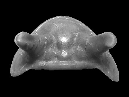
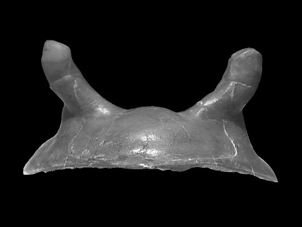
Average and large sized Asaphus (max : 12 cm). Occurence : Lower Ordovician,
Llanvirn. Body oval, rather broad and strongly convex.
- Cephalon : Rounded-triangular, strongly convex.
- Genal angles rather acute.
- Glabella pyriform, abruptly falling forward.
- Width / lenghth = 1.75
- Eyes large, on a high eyestalk basis, its heigth varying for various
species.
- More or less thick swelling oval in section in the basis of the eyes.
- Small knob behind the eyes (in the larger species), very much like
the knob of Asaphus kotlukowi but smoother.
- Palpebral lobes abruptly rising toward the rear part.
- Discontinuous terace lines on the anterior part of the glabella, absent
on the librigenæ.
- Thin pores and pits of sensor hairs on the surface.
- Thorax : Axial part narrower than the pleural one.
- Axial segments convex.
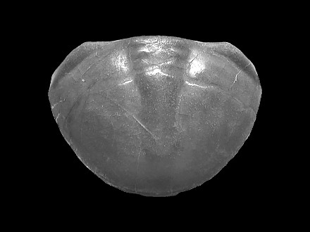
- Pygidium : Rounded-triangular.
- Slightly convex rachis, though clearly outlined.
- Segmentation seen on the lateral parts of the rachis.
- Thin pores and pits of sensor hairs on the surface.
- Width / lenghth = 1,6
|