Specimen description :
- Almost complet specimen.
- Lack of left genal spine.
- On matrix.
- Total size : 22 mm
- Lower Middle Ordovician - Llandeilian.
- Calzada de Calatrava.
- Ciudad Real, Spain.
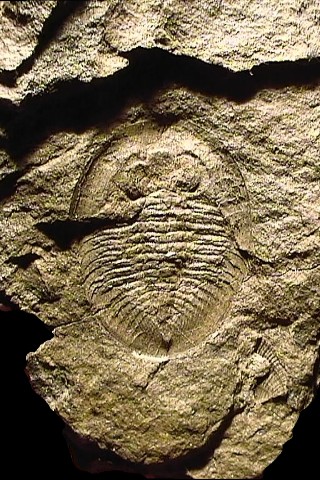
Dorsal view of the positive.
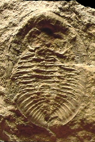
Ventral view of the negative.
Nobiliasaphus nobilis BARRANDE 1846
Nobiliasaphus nobilis is one the most frequently encountered asaphids of the Armorican Llanvirn (Brittany, France). It can be found in the Spanish Ordovician, as this is the case for the first specimen of that page. Even if my pictures don't show it that well, this is odd to remark that the 2 specimens do have exactly the same delicate golden color :)
|
Specimen description :
|
|
|
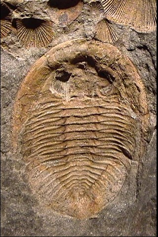
Dorsal view of the positive.
|
Ventral view of the negative.
|
Diagnosis :
|
Order
|
Asaphida
|
SALTER 1864
|
Order including 1/5 of all the species of Trilobites,
it mostly regroup librostoms of various morphology, in which the most advanced
families do present a ventral median suture early visible in the ontogeny
( "asaphoïd" protaspid larvæ).
Occurrence : Middle-Upper Cambrian boundary to upper Ordovician-lower Silurian.
|
|
Superfamily |
Asaphoidea |
BURMEISTER 1843 |
Occurrence: Middle Cambrian to Ordovician.
|
|
Family
|
Asaphidæ |
BURMEISTER 1843 |
Family tends toward loss of apparent segmentation of cephalon and pygidium, obsolescence of axial furrows and deep notching of posterior margin of hypostoma.
|
|
Subfamily |
Asaphinæ |
BURMEISTER 1843 |
|
|
Genus |
Nobiliasaphus |
PRIBYL & VANEK 1965 |
|
|
Species |
nobilis |
BARRANDE 1846
|
|
Complementary pictures :
 |
|
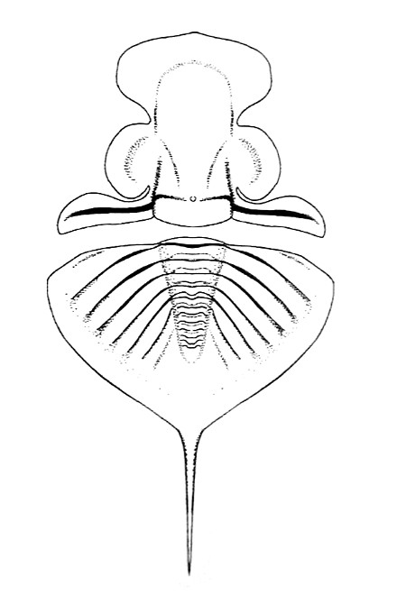
Synthetogram of the cephalon (D'après P. Lebrun,
2002)
|
Specimen of Brittany, France :
Trilobite found in the synclinorium of Laval.
 |
 |
|
ventral view of the positive part.
|
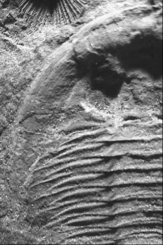
Zoom on the terrace lines of the cephalic border.
|
Diagnosis of Nobiliasaphus delessei (DUFET 1875) :
In Brittany, Nobiliasaphus delessei (DUFET 1875) can be collected in the Abereidian of the formation d'Angers.
| Species |
delessei |
DUFET (1875)
|
|
So, the important differential diagnostic point is the lack of median marginal pygidial spine for Nobiliasaphus nobilis.
 |
|