Description du spécimen
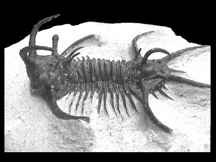
- Trilobite standing prone on matrix.
- Almost complete except for some tip of pleural spines.
- Exceptionnal preparation work.
- Total size : 70 mm.
- Devonian.
- Zerg, Morocco.
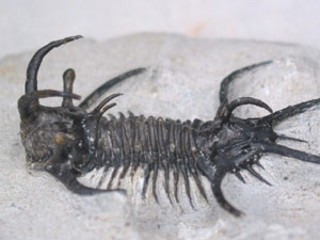
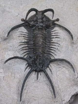
Dorsal view.
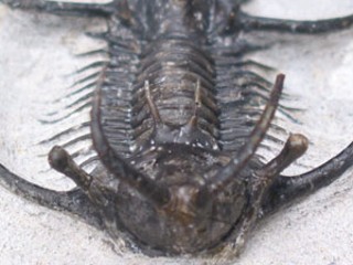
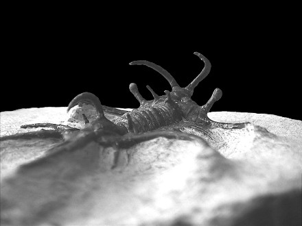
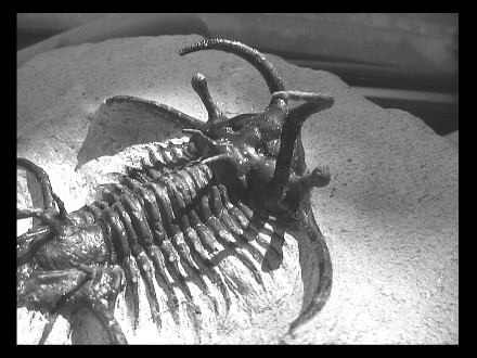
Up : frontal view. Down : Lateral view.
Ceratarges sp. n°1
Here is one of the breathtaking specimen of my collection. This beauty is due to Meredith Aaronson of Sahara Overland, whom Lab made a real high quality preparation work, where each of the spines could be set up free. The result is worth the price. Sadly, as I do lack a reference paper on Moroccan Lichids, I can't precisely ID this Ceratarges.
You can compare it to the second specimen I do present on the site. Some of the main differences here are the lack of secondary spines on the pygidial marginal spines, in contrast of median pairs of occipital spines and of a median pygidial spine. The occipital ring is more protruded, as the pygidial axis.
|
|
Description du spécimen
|
|
|
Dorsal view.
|
Up : frontal view. Down : Lateral view.
|
|
Diagnosis :
|
Order |
Lichida |
MOORE 1959 |
Typically spiny trilobites with densely granulate or tuberculate exoskeletons. Occurrence: Cambrian to Devonian (Frasnian)
|
|
Superfamily |
Lichoidea |
(sensu FORTEY 1991) |
Medium to large trilobites. Typically, surface sculpturing involves two size classes of granules or tubercles.
|
|
Family |
Lichidæ |
HAWLE & CORDA 1847 |
|
| Subfamily | Ceratarginæ | TRIPP 1957 |
|
| Genus | Ceratarges | GÜRICH 1901 |
|
| Species | sp. | to determine. (lack of references) |
Complementary pictures :
 |
 |
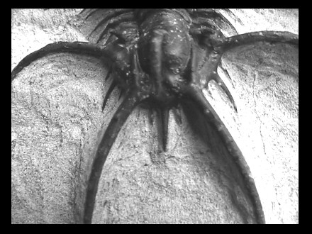
| Frontal view of the cephalon | Right postero lateral view of the specimen |
 |
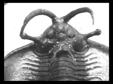 |
| Right postero lateral view of the cephalon | Dorsal view of the cephalon |
 |
 |
| Marginal spines of the cephalon, with an alternance in size. | Dorsal view of the thorax |
 |
 |
|
Median and adaxial pygidial spines.
|
Left lateral view of the specimen.
|