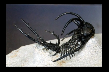
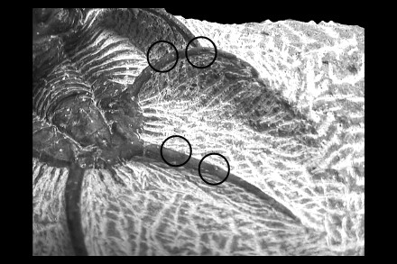
The main differences with the other specimen are the presence of secondary
spines on the pygidial marginal spines and the lack of occipital and pygidial
median and adaxial spines.
|
Order
|
Lichida
|
MOORE 1959
|
Typically spiny trilobites with densely granulate or tuberculate exoskeletons.
Occurrence: Cambrian to Devonian (Frasnian)
- Cephalon : Opisthoparian sutures.
- Glabella broad, large, extending to anterior border.
- Glabellar lobation simple (Dameselloidea & Odontopleuroidea) to complex
with fused lateral and glabellar lobes (Lichoidea).
- Eyes typically present, holochroal, usually not large.
- Conterminant hypostome.
- Thorax : Variable morphology.
- 8-13 segments usually spine-tipped, sometimes with distinctive spines
(e.g., Odontopleuroidea).
- Pygidium : Typically isopygous to macropygous, but sometimes
short (e.g., Odontopleuroidea).
- Often longer than wide.
- Often with 3 pairs of furrowed pleurae, typically ending in spinose
tips.
|
|
Superfamily
|
Lichoidea
|
(sensu FORTEY 1991)
|
Medium to large trilobites. Typically, surface sculpturing involves
two size classes of granules or tubercles.
- Cephalon : Opisthoparian sutures.
- Glabella widening close to the occipital ring, extending to anterior
border, with unique complex structure (lateral glabellar and occipital
lobes often fused with each other and with cranidium).
- L1a, L1b (subdivision of the preoccipital lobes) and bullar lobes
flanking the median glabellar lobe.
- Thorax: 10 - 11 segments.
- Pleurae initially horizontal, bend retrograde at fulcrum.
- Pleurae ending in free points.
- Pygidium: Large, usually flattened.
- Often with 3 pleural pairs of leaflike or spinose structures.
|
|
Family
|
Lichidæ
|
HAWLE &
CORDA 1847 |
- Cephalon : L1a, L2a and bullar lobes distinct, but sometimes
partially or totally fused with each other or with the adjacent part
of the fixigenae.
- Lack of preglabellar furrow in front of bullar lobe.
- Anterior branches of the facial sutures parallel or slightly convergent
anteriorly.
- No eye ridges.
|
| Subfamily |
Ceratarginæ |
TRIPP 1957 |
- Cephalon : Bicomposite lateral glabellar lobes bounded at back
by posterior lateral furrows (except in some species of Hemiarges).
- Axial furrows usually obsolete behind bicomposite lobes.
- Occipital lobes fused with basal lateral glabellar lobes (except in
Trochurus and Dicranogmus).
- Hypostoma with posterior margin not markedly indented. Middle body
circumscribed, with small posterior lateral lobes.
- Pygidium : Axis extended to posterior border or margin by narrow
ridge.
- Posterior bands of 1st and 2nd pleuræ narrower and more swollen
than anterior bands.
- Posterior pair of pleuræ unfurrowed.
|
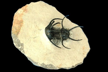
| Genus |
Ceratarges |
GÜRICH
1901 |
- Cephalon : Glabellar furrows faintly impressed.
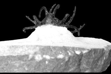
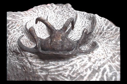
- Librigenal spines long and slender, forwardly placed.
- Long spines curving upward and backward on frontal lobe of glabella
and pygidium.
- Pygidium : Marginal spines.
|
| Species |
sp. |
|
to determine. (lack of references) |
Remark : better quality pictures to come soon.